Hip Joint
The hip joint is the largest weight-bearing joint in the human body. It is also referred to as a ball and socket joint and is surrounded by muscles, ligaments, and tendons. The thigh bone or femur and the pelvis join to form the hip joint.
Any injury or disease of the hip will adversely affect the joint's range of motion and ability to bear weight.
The hip joint is made up of the following:
- Bones and joints
- Ligaments of the joint capsule
- Muscles and tendons
- Nerves and blood vessels that supply the bones and muscles of the hip
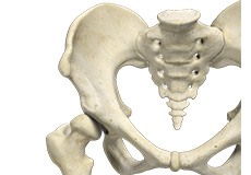
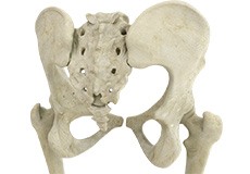
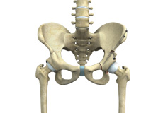
Bones and Joints
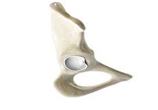
The hip joint is the junction where the hip joins the leg to the trunk of the body. It is comprised of two bones: the thigh bone or femur and the pelvis which is made up of three bones called ilium, ischium, and pubis. The ball of the hip joint is made by the femoral head while the socket is formed by the acetabulum. The Acetabulum is a deep, circular socket formed on the outer edge of the pelvis by the union of three bones: ilium, ischium, and pubis. The lower part of the ilium is attached by the pubis while the ischium is considerably behind the pubis. The stability of the hip is provided by the joint capsule or acetabulum and the muscles and ligaments which surround and support the hip joint.
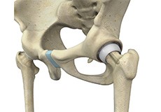
The head of the femur rotates and glides within the acetabulum. A fibrocartilagenous lining called the labrum is attached to the acetabulum and further increases the depth of the socket.
The femur or thigh bone is one of the longest bones in the human body. The upper part of the thigh bone consists of the femoral head, femoral neck, and greater and lesser trochanters. The head of the femur joins the pelvis (acetabulum) to form the hip joint. Next, to the femoral neck, there are two protrusions known as greater and lesser trochanters which serve as sites of muscle attachment.
Articular cartilage is the thin, tough, flexible, and slippery surface lubricated by synovial fluid that covers the weight-bearing bones of the body. It enables smooth movements of the bones and reduces friction.
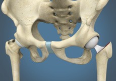
Ligaments
Ligaments are fibrous structures that connect bones to other bones. The hip joint is encircled with ligaments to provide stability to the hip by forming a dense and fibrous structure around the joint capsule. The ligaments adjoining the hip joint include:
Iliofemoral ligament: This is a Y-shaped ligament that connects the pelvis to the femoral head at the front of the joint. It helps in limiting the over-extension of the hip.
Pubofemoral ligament: This is a triangular shaped ligament that extends between the upper portion of the pubis and the iliofemoral ligament. It attaches the pubis to the femoral head.
Ischiofemoral ligament: This is a group of strong fibers that arise from the ischium behind the acetabulum and merge with the fibers of the joint capsule.
Ligamentum teres: This is a small ligament that extends from the tip of the femoral head to the acetabulum. Although it has no role in hip movement, it does have a small artery within that supplies blood to a part of the femoral head.
Acetabular labrum: The labrum is a fibrous cartilage ring which lines the acetabular socket. It deepens the cavity, increasing the stability and strength of the hip joint.
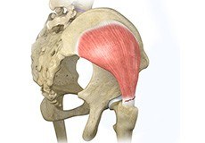
Muscles and Tendons
A long tendon called the iliotibial band runs along the femur from the hip to the knee and serves as an attachment site for several hip muscles including the following:
Gluteals: These are the muscles that form the buttocks. There are three muscles (gluteus minimus, gluteus maximus, and gluteus medius) that attach to the back of the pelvis and insert into the greater trochanter of the femur.
Adductors: These muscles are located in the thigh which helps in adduction, the action of pulling the leg back towards the midline.
Iliopsoas: This muscle is located in front of the hip joint and provides flexion. It is a deep muscle that originates from the lower back and pelvis and extends up to the inside surface of the upper part of the femur.
Rectus femoris: This is the largest band of muscles located in front of the thigh. They also are hip flexors.
Hamstring muscles: These begin at the bottom of the pelvis and run down the back of the thigh. Because they cross the back of the hip joint, they help in extension of the hip by pulling it backward.
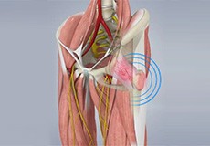
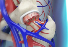
Nerves and Arteries
Nerves of the hip transfer signals from the brain to the muscles to aid in hip movement. They also carry the sensory signals such as touch, pain, and temperature back to the brain.
The main nerves in the hip region include the femoral nerve in the front of the femur and the sciatic nerve at the back. The hip is also supplied by a smaller nerve known as the obturator nerve.
In addition to these nerves, there are blood vessels that supply blood to the lower limbs. The femoral artery, one of the largest arteries in the body, arises deep in the pelvis and can be felt in front of the upper thigh.
Hip Movements
All of the anatomical parts of the hip work together to enable various hip movements. Hip movements include flexion, extension, abduction, adduction, circumduction, and hip rotation.

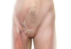
Hip Adductor Injuries
A hip adductor injury is also called a groin strain or groin tear and involves any of the adductor muscles. These injuries are common among athletes and those who play hockey, football, soccer, basketball, tennis, baseball, etc.
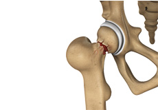
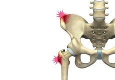
Stress Fractures of the Hip
Stress fractures of the hip are a break in the upper part of the thigh bone (femur) that fits into the socket of the hip joint. It can occur in any part of the hip, however, it mostly occurs just below the ball of the ball-and-socket hip joint called the femoral neck.
Avulsion Fractures of the Pelvis
Avulsion fractures of the pelvis is an injury that occurs when a tendon or ligament pulls off a piece of bone from the hip. This results in a part of the pelvic (hip) bone breaking away from the main part of the bone.
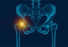
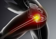
Hip Injury
The hip joint is one of the most important and flexible joints in the human body which allows us to walk, run, bend and perform physical activities. It is a ball (femoral head) and socket joint formed between the hip bone and femur (thighbone). It is surrounded by strong muscles and tough ligaments that prevent its dislocation.
Periprosthetic Hip Fractures
Hip replacement is a surgical procedure in which the damaged cartilage and bone are removed from the hip joint and replaced with artificial components. Any resulting fractures or breaks in the bone around the implant are called periprosthetic hip fractures. They usually occur around the stem of the implant and sometimes to the socket (acetabulum).
Gluteus Tendon Tear
The gluteal muscles (situated in the buttocks) are necessary for the stability and movement of the hip joints. The tendons of two gluteal muscles (gluteus medius and gluteal minimus) are attached at the outer hip region and are often called the “rotator cuff of the hip.” These tendons may be subject to injury or tearing due to various reasons.
Snapping Hip Syndrome
Snapping hip syndrome is a condition in which you hear or feel a snapping sound in the hip when you swing your legs, run, walk or get up from a chair. The sound can be experienced in the back, front or side of the hip.
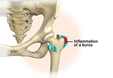
Hip Bursitis
Hip bursitis is a painful condition caused by the inflammation of a bursa in the hip. Bursae are fluid-filled sacs present in the joints between bone and soft tissue to reduce friction and provide cushioning during movement.
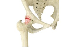
Femoroacetabular Impingement
Femoroacetabular impingement (FAI) is a condition characterized by excessive friction in the hip joint from the presence of bony irregularities. These cause pain and decreased range of hip motion.
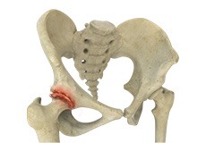
Avascular Necrosis
Avascular necrosis, also called osteonecrosis, is a condition in which bone death occurs because of inadequate blood supply to it. Lack of blood flow may occur when there is a fracture in the bone or a joint dislocation that may damage nearby blood vessels. Hip joint is most commonly affected; however, the knee and shoulder may also be involved.
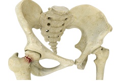
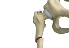
Hip Fracture
A hip fracture is a break that occurs near the hip in the upper part of the femur or thighbone. The thighbone has two bony processes on the upper part - the greater and lesser trochanters. The lesser trochanter projects from the base of the femoral neck on the back of the thighbone.
Hip Dislocation
The hip joint is a “ball and socket” joint. The “ball” is the head of the femur or thighbone, and the “socket” is the cup-shaped acetabulum. The joint is surrounded by muscles, ligaments, and tendons that support and hold the bones of the joint in place. Hip dislocation occurs when the head of the femur moves out of the socket.
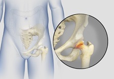
Hip Labral Tear
A hip labral tear is an injury to the labrum, the cartilage that surrounds the outside rim of your hip joint socket.
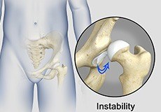
Hip Instability
Injury or damage to these structures can lead to a condition called hip instability when the joint becomes unstable. Hip instability can be traumatic or atraumatic. Traumatic instability can be caused by injuries from sports or motor vehicle accidents.
Hip Groin Disorders
Hip and groin disorders are more common in athletes. They are caused by rapid acceleration and deceleration motion. The rehabilitation time for hip and groin injuries is longer than most other injuries, therefore, early and accurate diagnosis is essential.
Subtrochanteric Hip Fracture
A hip fracture is a break that occurs near the hip in the upper part of the femur or thighbone. The thighbone has two bony processes on the upper part - the greater and lesser trochanters. The lesser trochanter projects from the base of the femoral neck on the back of the thighbone.
Hip Abductor Tears
Hip abductors are a major group of muscles found in the buttocks. It includes the gluteus maximus, gluteus medius, gluteus minimus, and tensor fascia lata muscles.
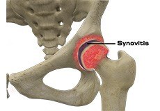
Hip Synovitis
Hip synovitis, also called transient hip synovitis or toxic synovitis, is a condition characterized by inflammation of the synovial tissues that surround the hip joint. It is the most common cause for sudden hip pain that occurs in young children between the age of 2 and 9. It affects boys more commonly than girls and is most of the time limited to only one side of the hip.
Developmental Dysplasia
Developmental dysplasia of the hip (DDH) or hip dysplasia is a condition that is seen in infants and young children because of developmental problems in the hip joint. The femur (thighbone) partially or completely slips out of the hip socket leading to dislocation at the hip joint. It is most common in the first-born baby with a family history of the disorder.
Irritable Hip
Irritable hip, also known as acute transient synovitis, is a common disorder of childhood characterized by hip pain and limping. The term transient means that it does not last long. It usually occurs before puberty and affects only one hip. Boys aged between 4 to 10 years are most often affected.
Hip Tendonitis
Tendons are strong connective tissue structures that connect muscle to bone. Hip tendonitis is a condition associated with degeneration of the hip tendons. This condition is mainly caused due to strain on the tendons which may occur due to overuse or biomechanical problems.
Hip Pointer
The hip joint consists of the femur (thighbone) and pelvic bone, which is made up of the fusion of three bones – the ischium, pubis, and ilium. The femur has two boney prominences close to the hip joint – the greater and lesser trochanters. Hip pointer is an injury or bruise to the iliac crest (curved upper border of the ilium) or greater trochanter, or the surrounding muscles or tissues.
Transient Osteoporosis of the Hip
Transient osteoporosis of the hip is a rare condition that causes temporary bone loss in the upper region of the thighbone (femur). It occurs most often in young or middle-aged men of the age groups 30 to 60, and women in their later stages of pregnancy or early postpartum (following childbirth). It is characterized by abrupt onset of pain that increases with activity.
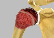
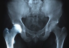
Osteoarthritis of the Hip
Osteoarthritis, also called degenerative joint disease, is the most common form of arthritis. It occurs most often in the elderly. This disease affects the tissue covering the ends of bones in a joint called cartilage. In osteoarthritis, the cartilage becomes damaged and worn out, causing pain, swelling, stiffness and restricted movement in the affected joint.
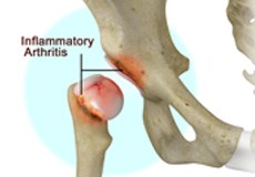
Inflammatory Arthritis of the Hip
The inflammation of the joints is referred to as arthritis. Inflammation arises when the smooth lining called cartilage at the ends of bones wears away. In some cases, the inflammation is caused when the lining of the joint becomes inflamed as part of an underlying systemic disease. These conditions are referred to as inflammatory arthritis.
Groin Injuries in Athletes
Groin injuries are injuries sustained by athletes during sports activity. Groin injuries comprise about 2 to 5 percent of all sports injuries. The most common kind of groin injury is a groin strain or a pulled groin muscle. Typical groin injuries vary based on athlete’s gender, age, and sports, with most common groin injuries occurring in certain sports such as football with a prevalence rate as high as 12-16%.
Periprosthetic Hip Infection
A very small percentage of patients (less than 1%) who undergo hip replacement may develop an infection around the hip joint following surgery. This infection is called a periprosthetic hip infection.
Hamstring Injuries
The hamstring is a group of three muscles that run along the back of the thigh from the hip to the knee. Hamstring injuries occur when these muscles are strained or pulled. They are common in dancers and athletes of all sorts including runners and those who play football, soccer, basketball, tennis, etc.
Mini-Posterior Hip Replacement
Mini-posterior hip replacement is a surgical procedure used to replace your damaged hip with synthetic parts inserted through a small incision made at the back of the hip.
Posterior Hip Replacement
Posterior hip replacement is a minimally invasive hip surgery performed to replace the hip joint. It is also referred to as muscle sparing surgery because no muscles are cut to access the hip joint, enabling a quicker return to normal activity.
Hip Fracture ORIF
A hip fracture is a break that occurs near the hip in the upper part of the femur or thighbone. The thighbone has two bony processes on the upper part - the greater and lesser trochanters. Open reduction and internal fixation (ORIF) is a surgical technique employed for the treatment of a fracture to restore normal anatomy and improve range of motion and function.
Outpatient Anterior Approach Hip Replacement
With improved technology and advances in anesthesia and pain control, hip replacement surgery has evolved and is now being offered in an outpatient setting.
Correction of a Failed Hip Replacement
Reoperation of a total hip replacement to resolve a painful hip condition arising out of a damaged or worn out prosthesis (artificial hip joint) is known as correction of a failed hip replacement. During this corrective surgery, a partial or complete exchange of the prostheses that were implanted during the original surgery is done.
Correction of a Painful Hip Replacement
Reoperation of a total hip replacement to resolve a painful hip condition arising out of a damaged or worn out prosthesis (artificial hip joint) is known as correction of a painful hip replacement. During this corrective surgery, a partial or complete exchange of the prostheses that were implanted during the original surgery is done.
Correction of a Loose Hip Replacement
Reoperation of a total hip replacement to resolve a painful hip condition and loss of motion due to a loosened prosthesis (artificial hip joint) is known as correction of a loose hip replacement. This loosening occurs due to wear and tear of the implant surfaces and subsequent weakening of the surrounding bone. During this corrective surgery, a partial or complete exchange of the prostheses that were implanted during the original surgery is done.
Outpatient Hip Replacement
Hip replacement surgery is one of the most common orthopedic surgeries performed. It involves the replacement of the damaged hip bone (ball shaped upper end of the femur) with a ceramic ball attached to a metal stem that is fixed into the femur and placing a new cup with a special liner in the pelvis.
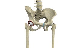
Hip Resurfacing
The hip joint is also known as a ball and socket joint, where the ball (femoral head) of the thigh bone fits into the socket (acetabulum) of the pelvic bone.
Hip Hemiarthroplasty
Hip hemiarthroplasty is a surgical technique employed to treat hip fractures. In this procedure, only one half (ball section) of the hip joint is substituted by a metal prosthesis.
Hip Distraction
The hip joint is one of the most important and flexible joints in the human body which allows us to walk, run, bend and perform physical activities. It is a ball (femoral head) and socket joint formed between the hip bone and femur (thigh bone). The hip joint is surrounded by strong muscles and tough ligaments that prevent dislocation of the hip.
Hip Preservation Surgery
The hip is a ball and socket joint comprising of the femur (thigh bone) and the pelvic bone. The head of the femur (ball) articulates with a cavity (socket) called the acetabulum in the pelvic bone. To facilitate the smooth and frictionless movement of the hip joint, the articulating surfaces of the femur head and acetabulum are covered by spongy articular cartilage.
Hip Fracture Surgery
Hip fractures involve a break that occurs near the hip in the upper part of the femur or thigh bone. The thigh bone has two bony processes on the upper part - the greater and lesser trochanters. The lesser trochanter projects from the base of the femoral neck on the back of the thigh bone. Hip fractures can occur either due to a break in the femoral neck, in the area between the greater and lesser trochanter or below the lesser trochanter.
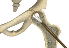
Core Decompression for Avascular Necrosis of the Hip
The hip joint is a ball and socket joint, where the head of the thighbone (femur) articulates with the cavity (acetabulum) of the pelvic bone. Sickle cell disease, a group of disorders that affect the hemoglobin or oxygen-carrying component of blood, causes avascular necrosis or the death of bone tissue in the hip due to lack of blood supply.
Surgical Release of Iliopsoas Tendon
Surgical release of the iliopsoas tendon is a procedure that involves the excision or cutting of the iliopsoas tendon in the hip to reduce pain and improve range of motion.
Physical Therapy for Hip
Physical therapy is an exercise program that helps you to improve movement, relieve pain, encourage blood flow for faster healing, and restore your physical function and fitness level. The main aim of physical therapy is to make your daily activities, such as walking, getting in and out of bed and climbing stairs, easier.
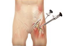
Hip Arthroscopy
Arthroscopy, also referred to as keyhole or minimally invasive surgery, is a procedure in which an arthroscope is inserted into a joint to check for any damage and repair it simultaneously.
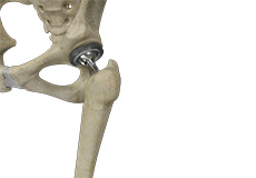
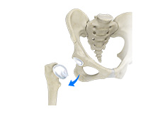
Total Hip Replacement
Total hip replacement is a surgical procedure in which the damaged cartilage and bone is removed from the hip joint and replaced with artificial components.
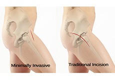
Minimally Invasive Total Hip Replacement
Surgery may be recommended in patients with severe cartilage damage and if conservative treatment options such as anti-inflammatory medications and physical therapy do not relieve the symptoms.
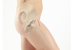
Anterior Hip Replacement
Anterior hip replacement surgery is performed under general anesthesia or regional anesthesia. You will lie down on your back, on a special operating table that enables your surgeon to perform the surgery from the front of the hip. Your surgeon may use fluoroscopic imaging during the surgery to ensure the accuracy of component positioning and to minimize leg length inequality.
Revision Hip Replacement
During total hip replacement, the damaged cartilage and bone are removed from the hip joint and replaced with artificial components. At times, hip replacement implants can wear out for various reasons and may need to be replaced with the help of a surgical procedure known as revision hip replacement surgery.
Computer-assisted Hip Replacement
Computer-assisted hip replacement is an image-guided, minimally invasive surgical procedure to replace your diseased or damaged hip with an artificial device using the assistance of computer software. The system creates and displays images and provides information that aids your surgeon at various stages of the procedure to improve accuracy and results.
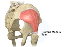
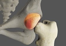
Gluteus Medius Tear
A gluteus medius tear is the partial or complete rupture of the gluteus medius muscle due to severe muscle strain. Gluteus medius tears often occur at the tendinous attachment to the greater trochanter of the femur bone.
Hip Trauma Reconstruction
Hip trauma is an injury in the hip due to the impact caused by incidents such as a car accident or a hard fall. The injury can be a bone break or dislocation or both.
Slipped Capital Femoral Epiphysis
Slipped capital femoral epiphysis (SCFE) is an unusual disorder of the hip where the ball at the upper end of the thighbone (femur) slips in a backward direction. This is caused due to weakness of the growth plate. This condition is commonly caused during accelerated growth periods such as the onset of puberty.
Computer-Navigated Total Hip Replacement
For a successful total hip replacement, accurate positioning of the implants is crucial to accomplish a good clinical outcome. Computer-navigated total hip replacement is an advanced technology developed to provide more accurate positioning of an implant. Hip replacement through computer navigation provides information and guidance to the surgeon for precise positioning of implants.
Direct Superior Hip Replacement
Direct superior hip replacement is a minimally invasive surgery that provides your surgeon access to the hip joint with minimum displacement or damage to the surrounding muscles, tendons, and ligaments.
Hip Reconstruction
Hip reconstruction is a surgery to repair or replace a damaged hip joint that causes pain and limits your movement.

Activities After Hip Replacement
Hip replacement is a surgery performed to replace parts of a diseased hip joint with a prosthesis. The goal of hip replacement is to eliminate pain and enable you to return to your normal activities. You can help in the recovery and improve the outcomes of the procedure by following certain precautions and changing the way you carry out your daily activities.
Physical Examination of the Hip
Physical examination of the hip is the key component in diagnosing the cause of your hip pain or hip pathology.