Knee Anatomy
The knee is a complex joint made up of different structures - bones, tendons, ligaments, and muscles. They all work together to maintain the knee’s normal function and provide stability to the knee during movement.
Having a well-functioning healthy knee is essential for our mobility and ability to participate in various activities. Understanding the anatomy of the knee enhances your ability to discuss and choose the right treatment procedure for knee problems with your doctor.
Bones of the Knee
The knee is a hinge joint made up of two bones, the thighbone (femur) and shinbone (tibia). There are two round knobs at the end of the femur called femoral condyles that articulate with the flat surface of the tibia called the tibial plateau. The tibial plateau on the inside of the leg is called the medial tibial plateau and on the outside of the leg, the lateral tibial plateau.
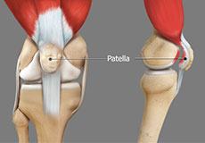
The two femoral condyles form a groove on the front (anterior) side of the knee called the patellofemoral groove. A small bone called the patella sits in this groove and forms the kneecap. It acts as a shield and protects the knee joint from direct trauma.
A fourth bone called the fibula is the other bone of the lower leg. This forms a small joint with the tibia. This joint has very little movement and is not considered a part of the main joint of the knee.
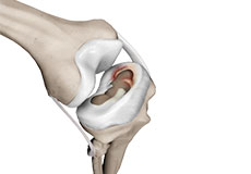
Articular Cartilage and Menisci of the Knee
Movement of the bones causes friction between the articulating surfaces. To reduce this friction, all articulating surfaces involved in the movement are covered with a white, shiny, slippery layer called articular cartilage. The articulating surface of the femoral condyles, tibial plateaus and the back of the patella are covered with this cartilage. The cartilage provides a smooth surface that facilitates easy movement.
To further reduce friction between the articulating surfaces of the bones, the knee joint is lined by a synovial membrane that produces a thick clear fluid called synovial fluid. This fluid lubricates and nourishes the cartilage and bones inside the joint capsule.
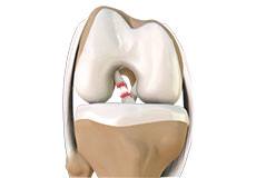
Within the knee joint, between the femur and tibia, are two C-shaped cartilaginous structures called menisci. Menisci function to provide stability to the knee by spreading the weight of the upper body across the whole surface of the tibial plateau. The menisci help in load-bearing i.e. it prevents the weight from concentrating onto a small area, which could damage the articular cartilage. The menisci also act as a cushion between the femur and tibia by absorbing the shock produced by activities such as walking, running and jumping.
Ligaments of the Knee
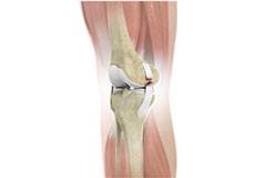
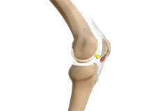
Ligaments are tough bands of tissue that connect one bone to another bone. The ligaments of the knee stabilize the knee joint. There are two important groups of ligaments that hold the bones of the knee joint together, collateral and cruciate ligaments.
Collateral ligaments are present on either side of the knee. They prevent the knee from moving too far during side to side motion. The collateral ligament on the inside is called the medial collateral ligament (MCL) and the collateral ligament on the outside is called the lateral collateral ligament (LCL).
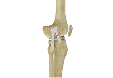
Cruciate ligaments, present inside the knee joint, control the back-and-forth motion of the knee. The cruciate ligament in the front of the knee is called anterior cruciate ligament (ACL) and the cruciate ligament in the back of the knee is called posterior cruciate ligament (PCL).
Muscles of the Knee
There are two major muscles in the knee - the quadriceps and the hamstrings, which enable movement of the knee joint. The quadriceps muscles are located in front of the thigh. When the quadriceps muscles contract, the knee straightens. The hamstrings are located at the back of the thigh. When the hamstring muscles contract, the knee bends.
Tendons of the Knee
A tendon is a tissue that attaches a muscle to a bone. The quadriceps muscles of the knee meet just above the patella and attach to it through a tendon called the quadriceps tendon. The patella further attaches to the tibia through a tendon called the patella tendon. The quadriceps muscle, quadriceps tendon, and patellar tendon all work together to straighten the knee. Similarly, the hamstring muscles at the back of the leg are attached to the knee joint with the hamstring tendon.
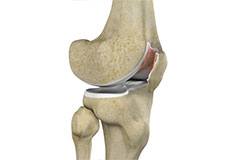
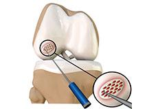
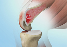
Chondromalacia Patella
Chondromalacia patella is a common condition characterized by softening, weakening and damage of the cartilage. The condition is most often seen in young athletes and older adults who have arthritis of the knee. It especially occurs in women.
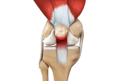
Jumper's Knee
Jumper’s knee, also known as patellar tendinitis, is inflammation of the patellar tendon that connects your kneecap (patella) to your shinbone. This tendon helps in the extension of the lower leg.
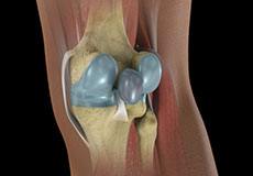
Baker's Cyst
The knee consists of a fluid called synovial fluid, which reduces the friction between the bones of the knee joint while you move your leg. Sometimes this fluid is produced in excess, resulting in its accumulation in the back of your knee. A Baker’s cyst or popliteal cyst is a fluid-filled swelling that develops into a lump behind the knee.
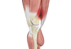
Iliotibial Band Syndrome
Iliotibial band syndrome is an overuse injury resulting from the inflammation of the iliotibial band. It occurs when the iliotibial band and the lower outside portion of the thighbone at the knee joint rub against each other.
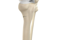
Fractures of the Tibia
The lower leg is made up of two long bones called the tibia and fibula that extend between the knee and ankle. The tibia or shinbone is the larger of the two bones. It bears most of the body’s weight and helps form the ankle joint and knee joint.
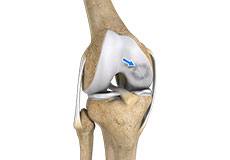
Osteochondritis Dissecans of the Knee
Osteochondritis dissecans is a joint condition in which a piece of cartilage, along with a thin layer of the bone separates from the end of the bone because of inadequate blood supply. The separated fragments are sometimes called “joint mice”.
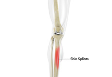
Shin Splints
Shin splints are pain and inflammation of the tendons, muscles and bone tissue along the tibia or shinbone (lower leg). It occurs because of vigorous physical activities such as exercise or sports. The condition is also referred to as medial tibial stress syndrome (MTSS).
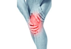
Knee Injury
Pain, swelling, and stiffness are the common symptoms of any damage or injury to the knee. If care is not taken during the initial phases of injury, it may lead to joint damage, which may end up destroying your knee.
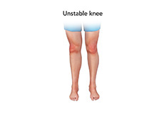
Unstable Knee
Damage to any of these supportive structures causes instability of the knee joint. An unstable knee can be caused by the sudden twisting of the knee, tears of the meniscus, ligament or capsule, osteoarthritis of the knee (wear and tear of the cushioning cartilage tissue between the bones) and sports injuries.
Knee Sprain
Knee sprain is a common injury that occurs from overstretching of the ligaments that support the knee joint. A knee sprain occurs when the knee ligaments are twisted or turned beyond its normal range, causing the ligaments to tear.

Knee Infection
Knee infection is a serious medical condition that needs immediate treatment. Infection may occur followed by a knee replacement surgery or trauma and is usually caused by bacteria. Infection may spread to the space of the knee joint or deep layers of your knee causing serious complications.
ACL Tears
The anterior cruciate ligament (ACL) is one of the major ligaments of the knee. It is located in the middle of the knee and runs from the femur (thighbone) to the tibia (shinbone). The ACL prevents the tibia from sliding out in front of the femur. Together with the posterior cruciate ligament (PCL), it provides rotational stability to the knee.
MCL Sprains
MCL sprains occur due to a sudden impact from the outside of your knee, most commonly while playing sports such as rugby and football. Rarely, the MCL can get injured when the knee gets twisted or following a quick change in direction./p>
Meniscal Injuries
Meniscal tears are among the commonest injuries to the knee joint. It can occur at any age but are more common in athletes involved in contact sports. The meniscus has no direct blood supply and for that reason, when there is an injury to the meniscus, healing is difficult.
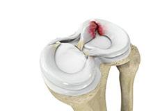
Meniscal Tears
A meniscal tear is a common knee injury in athletes, especially those involved in contact sports. A sudden bend or twist in your knee causes the meniscus to tear. Elderly people are more prone to degenerative meniscal tears as the cartilage wears out and weakens with age.

Ligament Injuries
The knee is a hinge joint made up of two bones, the thighbone (femur) and shinbone (tibia). Ligaments are tough bands of tissue that connect one bone to another bone. The ligaments of the knee stabilize the knee joint.
Knee Arthritis
The joint surface is covered by a smooth articular surface that allows pain-free movement in the joint. Arthritis is a general term covering numerous conditions where the joint surface or cartilage wears out. This surface can wear out for several reasons; often the definite cause is not known.
Patellar Dislocation/Patellofemoral Dislocation
Patellar dislocation occurs when the patella moves out of the patellofemoral groove, (trochlea) onto the bony head of the femur. If the kneecap partially comes out of the groove, it is called subluxation; if the kneecap completely comes out, it is called dislocation (luxation).
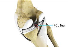
PCL Injuries
Posterior cruciate ligament (PCL), one of the four major ligaments of the knee, is situated at the back of the knee. It connects the thighbone (femur) to the shinbone (tibia). The PCL limits the backward motion of the shinbone.
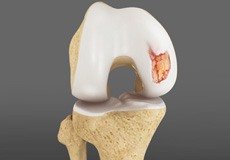
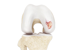
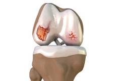
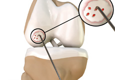
Chondral or Articular Cartilage Defects
The articular or hyaline cartilage is the tissue lining the surface of the two bones in the knee joint. Cartilage helps the bones move smoothly against each other and can withstand the weight of your body during activities such as running and jumping. Articular cartilage does not have a direct blood supply to it, so has less capacity to repair itself.
Patellar Instability
Any damage to the supporting ligaments may cause the patella to slip out of the groove either partially (subluxation) or completely (dislocation). This misalignment can damage the underlying soft structures such as muscles and ligaments that hold the kneecap in place.
Patellofemoral Instability
Patellofemoral instability means that the patella (kneecap) moves out of its normal pattern of alignment. This malalignment can damage the underlying soft structures such as muscles and ligaments that hold the knee in place.
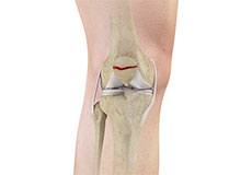
Patella Fracture
The kneecap or patella forms a part of the knee joint. It is present at the front of the knee, protecting the knee and providing attachment to various muscle groups of the thigh and leg. The undersurface of the kneecap and the lower end of the femur are coated with articular cartilage, which helps in smooth movement of the knee joint. A fracture in the kneecap is rare but common in adult males.
Recurrent Patella Dislocation
The patella (kneecap) is a small bone that shields your knee joint. It is present in front of your knee, on a groove called the trochlear groove that sits at the junction of the femur (thighbone) and tibia (shinbone). Articular cartilage presents below the patella and end of the femur cushion and helps the bones glide smoothly over each other when the legs move.
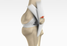
Quadriceps Tendon Rupture
The quadriceps tendon is a thick tissue located at the top of the kneecap. It works together with the quadriceps muscles to allow us to straighten our leg. The quadriceps muscles are the muscles located in front of the thigh.
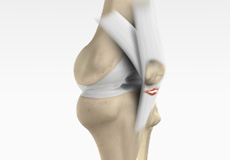
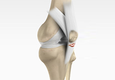
Patellar Tendon Rupture
The patellar tendon works together with the quadriceps muscle and the quadriceps tendon to allow your knee to straighten out. Patella tendon rupture is the rupture of the tendon that connects the patella (kneecap) to the top portion of the tibia (shinbone).
Lateral Meniscus Syndrome
Lateral meniscus syndrome is characterized by an injury caused by the tearing of the cartilage tissue or a rare case of a congenital abnormality called a discoid meniscus, which results in knee pain.
Osteonecrosis of the Knee
Osteonecrosis is a condition in which the death of a section of bone occurs because of lack of blood supply to it. It is one of the most common causes of knee pain in older women. Women over 60 years of age are commonly affected, three times more often than men.
Knee Angular Deformities
Angular deformities of the knee are variations in the normal growth pattern during early childhood and are common during childhood. The condition usually becomes more evident when your child is 2 to 3 years old and normally corrects itself by 7 or 8 years.
Periprosthetic Knee Fractures
Knee replacement, also called knee arthroplasty, is a surgical procedure in which the worn-out or damaged surfaces of the knee joint are removed and replaced with artificial implants. Any resulting fractures or breaks in the bone around the implant are called periprosthetic knee fractures.
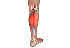
Medial Gastrocnemius Strain
A medial gastrocnemius strain (MGS), also sometimes called “tennis leg”, is an injury to the calf muscle in the back of the leg. It occurs when the calf muscle is stretched too far resulting in a partial or total tear or rupture within the muscle.
Articular Cartilage Injury
Articular or hyaline cartilage is the tissue lining the surface of the two bones in the knee joint. Cartilage helps the bones move smoothly against each other and can withstand the weight of the body during activities such as running and jumping. Articular cartilage does not have a direct blood supply to it so has little capacity to repair itself.
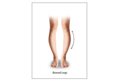
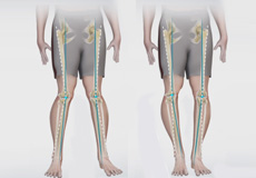
Bowed Legs
Bowed leg is a bony deformity resulting in outward curvature of one or both knees of the lower legs. It is commonly seen in toddlers and overweight adolescents.
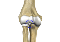
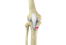
Loose Bodies in the Knee
Loose bodies are fragments of detached cartilage or bone inside the knee joint. These fragments may be free floating (unstable) or may be trapped (stable) within the joint. Depending on the severity, you may have one or more loose bodies in your knee joint.
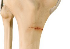
Knee Fracture
A fracture is a condition in which there is a break in the continuity of the bone. In younger individuals, these fractures are caused by high energy injuries, as from a motor vehicle accident. In older people, the most common cause is a weak and fragile bone.
Knee Osteoarthritis
Osteoarthritis also called degenerative joint disease, is the most common form of arthritis. It occurs most often in older people. This disease affects the tissue covering the ends of bones in a joint (cartilage).In a person with osteoarthritis, the cartilage becomes damaged and worn out causing pain, swelling, stiffness and restricted movement in the affected joint.
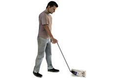
Knee Sports Injuries
Trauma is any injury caused during physical activity, motor vehicle accidents, electric shock, or other activities. Sports trauma or sports injuries refer to injuries caused while playing indoor or outdoor sports and exercising.
Patellar Tendinitis
Patellar tendinitis, also known as "jumper's knee", is an inflammation of the patellar tendon that connects your kneecap (patella) to your shinbone. This tendon helps in extension of the lower leg.
Women and ACL Injuries
The anterior cruciate ligament is one of the four major ligaments of the knee that connects the femur (thigh bone) to the tibia (shin bone) and helps stabilize the knee joint. Anterior cruciate ligament (ACL) injury is one of the common injuries of the knee.
Periprosthetic Knee Infection
A very small percentage of patients (less than 1%) who undergo knee replacement may develop an infection around the knee joint. This infection is called a periprosthetic knee infection.
Medial Meniscus Syndrome
Medial meniscal injuries are usually considered as either traumatic or degenerative. Whilst degenerate tears may present with a gradual history of increasing symptoms, traumatic injuries will usually occur as the knee is extended and rotated from a flexed position against resistance.
Multiligament Knee Injuries
Injury to more than one knee ligament is called a multiligament knee injury and may occur during sports or other physical activities.
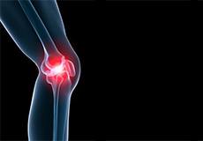
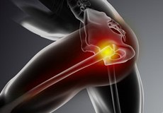
Knee Pain
Knee pain is a common condition affecting individuals of different age groups. It not only affects movement but also impacts your quality of life. An injury or disease of the knee joint or any structure surrounding the knee can result in knee pain. A precise diagnosis of the underlying cause is important to develop an appropriate treatment plan.
Anterior Knee Pain
Anterior knee pain is characterized by chronic pain over the front and center of the knee joint. It is common in athletes, active adolescents (especially girls) and overweight individuals. Anterior knee pain refers to various conditions, which include runner's knee or patellar tendinitis, and chondromalacia of the patella.
Runner's Knee
Patellofemoral pain syndrome also called runner’s knee refers to pain under and around your kneecap. Patellofemoral pain is associated with a number of medical conditions such as anterior knee pain syndrome, patellofemoral malalignment, and chondromalacia patella. Patellofemoral pain is a common complaint among runners, jumpers, and other athletes such as skiers, cyclists, and soccer players; thus the common name, runner’s knee.
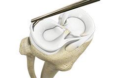
Partial Arthroscopic Meniscectomy
Partial arthroscopic meniscectomy is a procedure to remove the damaged part of a meniscus in the knee joint with the help an arthroscope. The meniscus is a C-shaped disc of cartilage between your thighbone and shinbone. There are 2 menisci in each knee. They act as shock absorbers and provide stability to the joint.
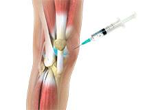
Combined Hyaluronic Therapy for the Knee
Combined hyaluronic therapy is the process of injecting hyaluronic acid (HA) along with platelet-rich plasma (PRP) into your knee to treat osteoarthritis.
Correction of a Failed Knee Replacement
Reoperation of a total knee replacement to resolve a painful knee condition and loss of motion arising out of a damaged or worn out prosthesis is known as correction of a failed knee replacement. This procedure involves a partial or complete exchange of the prostheses that were implanted during the original total knee replacement with new prostheses.
Correction of a Loose Knee Replacement
Reoperation of a total knee replacement to correct a loosened prosthesis as a result of wear and tear of the prosthetic joint surfaces is known as correction of a loose knee replacement. This procedure involves a complete or partial exchange of prostheses implanted during the original total knee replacement with new prostheses.
Correction of a Painful Knee Replacement
Reoperation of a total knee replacement to resolve a painful knee condition and loss of motion arising out of a damaged or worn out prosthesis is known as correction of a painful knee replacement. This procedure involves a complete or partial exchange of prostheses implanted during the original total knee replacement with new prostheses.
Knee Fracture Surgery
A knee fracture is a broken bone or a crack in or around the joint of the knee. This can involve the tibia (shin bone), the kneecap (patella), or femur (thighbone) where they connect with the knee.
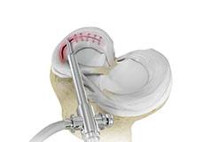
Arthroscopic Debridement
Arthroscopic debridement or a clean-up is a surgical procedure performed using an arthroscope. In this procedure, the cartilage or the bone that is damaged is removed using surgical instruments and the edges of the articular cartilage that are rough will be smoothened. A washout or joint lavage is done using a special tool to spray jets of fluid to wash and suck out to remove the remaining debris around the joint.
ConforMIS Knee
ConforMIS® has developed personalized knee resurfacing implants with unique advantages against traditional knee replacement options. To design a personalized implant for every individual, the surgeon will perform the CT scan of the affected knee of the patient.
Failed Meniscus Repair
Meniscal repair may be performed either by open surgery under direct vision or minimally invasively using an arthroscope, which is a thin tube fitted with a camera that can be inserted into the knee through a very small incision to locate and repair the damaged meniscus.
Painful or Failed Total Knee Replacement
Total knee replacement is a surgery employed to resurface knee joints damaged by arthritis, degeneration, or injury and replacing the damaged joints with a prosthesis (an artificial knee joint).
Meniscectomy
Meniscectomy is a surgical procedure indicated in individuals with torn meniscus where the conservative treatments are a failure to relieve the pain and other symptoms. Meniscectomy is recommended based on the ability of meniscus to heal, patient’s age, health status, and activity level.
Minimally Invasive Knee Joint Replacement
Total knee replacement is a very successful surgical treatment for knee arthritis. Over the years, minimally invasive knee replacement surgical techniques have been developed to lessen tissue trauma and improve patient outcomes.
Outpatient Unicondylar Knee Replacement
A unicondylar knee replacement, also known as unicompartmental or partial knee replacement, is a procedure to replace a portion of the damaged knee joint with a prosthetic implant to relieve pain and improve function of the knee joint.
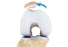
Partial Knee Resurfacing
Partial knee replacement is an alternative to total knee replacement in patients with arthritis on only one side of the knee. Partial knee replacement is a surgical procedure which involves resurfacing and replacement of only the diseased surface of the joint instead of the entire joint.
Patient Specific Knee Replacement
Patient Specific Knee Replacement is a newer technology in total knee replacement surgery. It is an advanced procedure using an individualized patient-specific knee implant for replacement of all three components of the knee.
Prior Meniscectomy
The menisci are two C-shaped cartilages that act as shock absorbers between the thigh and shin bones that articulate at the knee joint. They provide stability and lubrication to the joint as well as nutrition for the articular cartilage.
Unicondylar knee Replacement
Unicompartmental knee replacement is a minimally invasive surgery in which only the damaged compartment of the knee is replaced with an implant. It is also called a partial knee replacement. The knee can be divided into three compartments: patellofemoral, the compartment in front of the knee between the knee cap and thigh bone, medial compartment, on the inside portion of the knee, and lateral compartment which is the area on the outside portion of the knee joint.
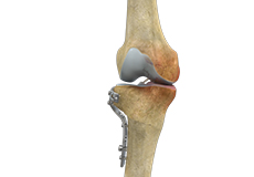
Periprosthetic Knee Fracture Fixation
Periprosthetic knee fracture fixation is a procedure performed to stabilize a fracture that occurs in the bone present around a knee prosthesis. The fracture may involve the lower part of the thighbone (femur), the kneecap (patella) or the upper part of the shinbone (tibia).
ORIF of the Knee Fracture
ORIF refers to open reduction and internal fixation. It is a surgical procedure employed for the treatment of a fracture not amenable to non-surgical conservative treatment.
Custom Knee Replacement
Custom Knee Replacement is an advanced surgical procedure in which the damaged knee joint is replaced by a customized implant, specifically designed to match the unique size and shape of each patient’s knee.
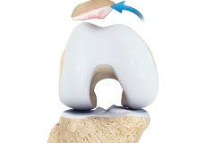
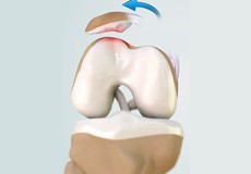
Chondroplasty
Chondroplasty is a surgical procedure to repair and reshape damaged cartilage in a joint. The procedure involves smoothing degenerative cartilage and trimming any unstable flaps of cartilage.
Pharmacological Interventions for Knee Injuries
The knee is a complex joint that consists of bone, cartilage, ligaments, and tendons, which help in joint movements. Knee problems may arise if any of these structures get injured by overuse, trauma or during sports activities. Pharmacological interventions include medicinal preparations such as pain-relieving capsules or injections.
Viscosupplementation
Viscosupplementation refers to the injection of a hyaluronan preparation into the joint. Hyaluronan is a natural substance present in the joint fluid that assists in lubrication. It allows the smooth movement of the cartilage-covered articulating surfaces of the joint.
Physical Therapy for Knee
Physical therapy is an exercise program that helps you to improve movement, relieve pain, encourage blood flow for faster healing, and restore your physical function and fitness level. It can be prescribed as an individual treatment program or combined with other treatments. It involves a combination of education, manual therapy, exercises and techniques such as water, heat, cold, electrical stimulation and ultrasound.
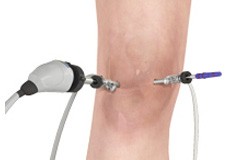
Knee Arthroscopy
Knee arthroscopy is a common surgical procedure performed using an arthroscope, a viewing instrument, to diagnose or treat a knee problem. It is a relatively safe procedure and you will usually be discharged from the hospital on the same day of surgery.
High Tibial Osteotomy
High tibial osteotomy is a surgical procedure performed to relieve pressure on the damaged site of an arthritic knee joint. It is usually performed in arthritic conditions affecting only one side of your knee and the aim is to take pressure off the damaged area and shift it to the other side of your knee with healthy cartilage.
Tibial Tubercle Osteotomy
Tibial tubercle osteotomy is a surgical procedure that is performed along with other procedures to treat patellar instability, patellofemoral pain, and osteoarthritis. The tibial tubercle transfer technique involves realignment of the tibial tubercle (a bump in the front of the shinbone) such that the kneecap (patella) traverses in the center of the femoral groove.
Unicompartmental/Partial Knee Replacement
Unicompartmental knee replacement is a minimally invasive surgery in which only the damaged compartment of the knee is replaced with an implant. It is also called a partial knee replacement.
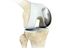
Patellofemoral Knee Replacement
Traditionally, arthritis in only one compartment of the knee is treated by partial knee replacement surgery. Patellofemoral knee replacement is a minimally invasive surgical option performed in the patellofemoral compartment only, preserving the knee parts not damaged by arthritis as well as the stabilizing anterior and posterior cruciate ligaments (ACL and PCL).
What is New in Knee Replacement
If you are considering knee replacement surgery, there are new developments under study which can help enhance the quality of life.
Computer Navigation for Total Knee Replacement
Computer navigation provides your surgeon with real-time 3-D images of your mapped knee and the surgical instruments during surgery. The data for the images is provided by infrared sensors fixed to the bones of the knee and surgical instruments. Their position is tracked by an infrared camera placed above the surgical table, which is connected to a computer.
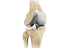
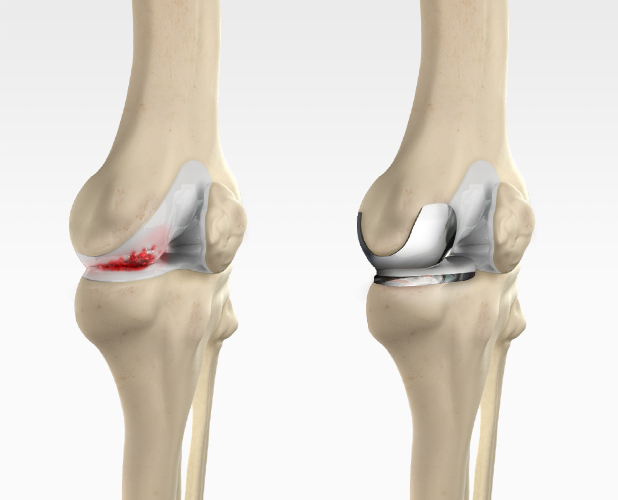
Total Knee Replacement
Total knee replacement, also called total knee arthroplasty, is a surgical procedure in which the worn out or damaged surfaces of the knee joint is removed and replaced with an artificial prosthesis.
Revision Knee Replacement
Revision knee replacement surgery involves replacing a part or all your previous knee prosthesis with a new prosthesis. Although total knee replacement surgery is successful, sometimes the procedure can fail due to various reasons and may require a second revision surgery.
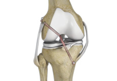
ACL Reconstruction
ACL reconstruction is a commonly performed surgical procedure. With recent advances in arthroscopic surgery, it can now be performed with minimal incision and low complication rates.
Outpatient Total Knee Replacement
Total knee replacement is the surgical treatment for knee arthritis, where the damaged knee is removed and replaced with an artificial knee implant. Traditionally performed as an inpatient procedure, total knee replacement surgery is now being conducted on an outpatient basis, allowing you to go home on the same day of the surgery.
Tricompartmental Knee Replacement
Tricompartmental knee replacement, also called total knee arthroplasty, is a surgical procedure in which the worn-out or damaged surfaces of the knee joint are removed and replaced with artificial parts.
Custom-fitted Total Knee Arthroplasty
Custom-fitted total knee arthroplasty is a newer more advanced technology in total knee replacement surgery that uses an individualized patient-specific knee implant for the replacement of all three components of the knee.
Signature Knees
Signature knee is a unique knee replacement procedure that is tailored to suit your individual anatomy. The technique utilizes magnetic resonance imaging (MRI) technology to create 3D images of your knee joint, which enables your doctor to efficiently plan your knee replacement surgery.
Bicompartmental Knee Resurfacing
Bicompartmental knee resurfacing is a less invasive surgical alternative to total knee replacement surgery, where instead of all the compartments being replaced only 2 of the 3 compartments of the knee damaged by arthritis are replaced with a prosthesis.
Subchondroplasty
Subchondroplasty is a minimally invasive procedure that is performed to specifically repair chronic BMLs by filling them with a bone substitute material. The bone substitute is then slowly resorbed and replaced with healthy bone, repairing the bone defect. Subchondroplasty also resolves the associated edema.
Partial Meniscectomy
Partial meniscectomy is a surgical procedure to remove the torn portion of the meniscus from the knee joint.
Meniscal Surgery
A meniscus tear is the commonest knee injury in athletes, especially those involved in contact sports. A sudden bend or twist in your knee can cause the meniscus to tear. This is a traumatic meniscal tear. The elderly are more prone to degenerative meniscal tears as the cartilage wears out and weakens with age.
Partial Lateral Knee Replacement
Partial lateral knee replacement is a surgery to replace only the lateral part of your damaged knee. It is also called unicompartmental knee replacement.
Partial Medial Knee Replacement
Partial medial knee replacement is a surgery to replace only the medial part of your damaged knee. It is also called unicompartmental knee replacement. The knee is one of the largest and complex joints in your body. The joint is connected to your thigh bones and bones of the lower leg by various ligaments.
Nonoperative Treatments for ACL Injuries
Nonoperative treatments may be recommended if your child has a minor ACL injury, is still growing and does not wish to immediately return to sports.
Non-Surgical Knee Treatments
The knee is a complex joint which consists of bone, cartilage, ligaments, and tendons that make joint movements easy and at the same time it is more susceptible to various kinds of injuries. Knee problems may arise if any of these structures get injured by overuse or suddenly during sports activities.
Physical Examination of the Knee
A complete physical examination of the knee is performed when you present to your doctor with a knee complaint. Both of your knees are examined and the results of the injured knee are compared to those of the healthy knee.
Pre-op and Post-op Knee Guidelines
Planning for your knee surgery prepares you for the operation and helps to ensure a smooth surgery and easier recovery. Here are certain pre-operative and post-operative guidelines which will help you prepare for knee surgery.
Am I a Candidate for Knee Surgery?
Arthritis of the knee can cause pain and stiffness, making regular activities such as walking and bending difficult. As arthritis progresses, conservative treatments tend to lose their efficacy and more definitive treatment should be considered. Knee replacement surgery involves replacing worn or damaged joints with implants to reduce pain and improve movement.
After Knee Replacement
After knee replacement surgery, once the anesthesia wears off, you will start to experience pain, for which your doctor will prescribe medication. You may have to remain in the hospital for a few days depending on your progress and overall health. Remember to get plenty of rest during this initial phase.
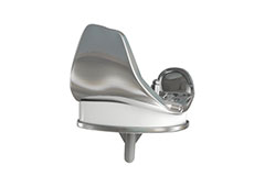
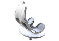
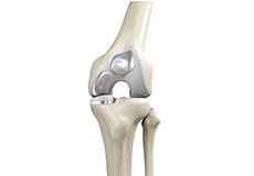
Knee Implants
Knee implants are artificial devices that form the essential parts of the knee during a knee replacement surgery. The knee implants vary by size, shape, and material. Implants are made of biocompatible materials that are accepted by the body without producing any rejection response.